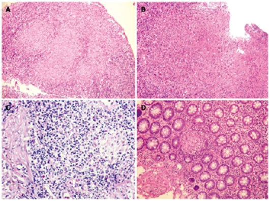
Figure 4.

Histological features. A: Confluent granulomas in inflammatory granulation tissue from ulcerated colonic mucosa of a patient with tuberculosis (TB) [Hematoxylin and eosin (HE), 100 ×]; B: Large granuloma in the ulcerated mucosa of a patient with TB (HE, 100 ×); C: Microgranuloma composed of a small aggregate of macrophages in a lymphoid follicle from the mucosa of a patient with Crohn's disease (CD) (HE, 400 ×); D: Small pericryptal granuloma in the colonic mucosa of a patient with CD (HE, 100 ×).
