
Figure 1.
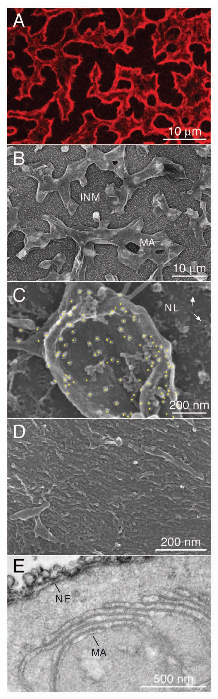
Expression of lamin B2 induces intranuclear membrane-like arrays. FLAG-tagged Xenopus lamin B2 was expressed in Xenopus oocytes by nuclear injection of plasmid DNA. (A) Immunofluorescence microscopy of a nuclear envelope spread of a FLAG-lamin B2 expressing oocyte (top view). FLAG-tagged lamin B2 (red) was detected with mAb M2 and a Cy3-conjugated secondary antibody. (B) feSEM image of a nuclear envelope spread of a lamin B2 expressing oocyte taken at low magnification. Lamin-induced intranuclear arrays (MA) are attached to the inner nuclear membrane (INM). The inner membrane is decorated by numerous pore baskets. (C) Immuno-feSEM detection of FLAG-tagged lamin B2 on the surface of an intranuclear array using mAb M2. Immuno-gold particles were identified in a backscatter image and marked by yellow spots. Gold particles are restricted to the surface of the intranuclear array while the nuclear lamina (NL) is not labeled. Arrows in the upper right point to nuclear pore baskets. (D) feSEM image of the surface of an intranuclear array taken at higher magnification. Individual lamin filaments cover the entire surface of the array. (E) TEM section of an isolated oocyte nucleus expressing lamin B2. Note the similarity between the nuclear envelope cisternae (NE) and the stacked arrays (MA).