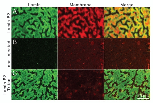
Figure 2.
Detection of lipids in lamin-induced intranuclear arrays. Nuclear envelope spreads of Xenopus oocytes were stained for membrane lipids with CM-DiI (Membrane). FLAG-tagged lamin B2 was detected by indirect immunofluorescence with mAb L7-8C6 and a FITC-conjugated secondary antibody (Lamin). Spreads were analyzed by confocal fluorescence microscopy. (A) Spreads of lamin B2 expressing oocytes, (B) spreads of non-injected control oocytes, (C) spreads of lamin B2 expressing oocytes that were treated with Triton X-100 prior to staining for lipids. The right parts show merged images. Red intensively fluorescing dots in (B and C) (Membrane) are due to small insoluble CM-DiI particles that stick to the samples. Size bar for all parts 10 µm.

