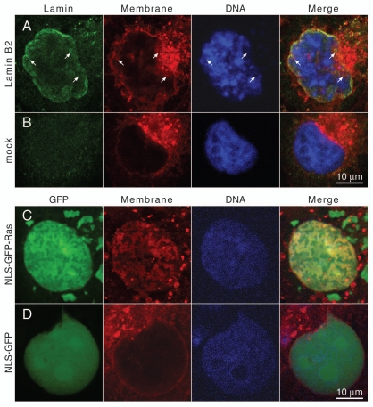
Figure 3.
Intranuclear arrays induced by expression of isoprenylated nuclear proteins in Cos-7 cells contain membrane lipids. (A) Cos-7 cells transiently transfected with FLAG-tagged lamin B2 or (B) mock transfected cells were stained for membrane lipids with CM-DiI (red) 24 hours after transfection, then fixed and permeabilized and stained for lamin B2 with mAb M2 and an FITC-conjugated secondary antibody (green) and for DNA with DAPI (blue). (C) Cos-7 cells transiently transfected with NLS-GFP-Ras or (D) NLS-GFP were stained with CM-DiI (red) 24 hours after transfection, they were then fixed and stained for DNA with DAPI (blue). FITC and GFP fluorescence was detected in the green channel. Samples were analyzed with a confocal fluorescence microscope. Mid-nuclear optical sections are shown. Arrows point to intranuclear membranes. The right parts show merged images of all three channels. Size bar for all parts 10 µm.

