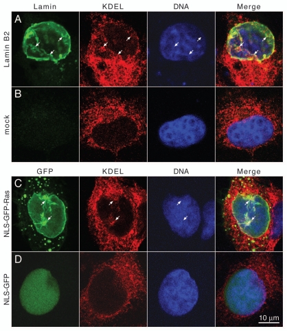
Figure 4.
Intranuclear membrane cisternae contain luminal ER proteins. (A) Cos-7 cells transiently transfected with FLAG-tagged lamin B2 or (B) mock transfected cells were fixed and stained for lamin B2 with polyclonal rabbit antibodies directed against the FLAG-epitope and an FITC-conjugated secondary antibody (green), for KDEL-containing ER proteins with mAb 10C3 and a Cy3-conjugated secondary antibody (red) and for DNA with DAPI (blue). (C) Cos-7 cells transiently transfected with NLS-GFP-Ras or (D) transfected with NLS-GFP were stained for KDEL proteins and for DNA as described above. FITC and GFP fluorescence was detected in the green channel. Cells were analyzed by confocal fluorescence microscopy. Mid-nuclear optical sections are shown. Arrows point to intranuclear membranes. The right parts show merged images of all three channels. Size bar for all parts 10 µm.

