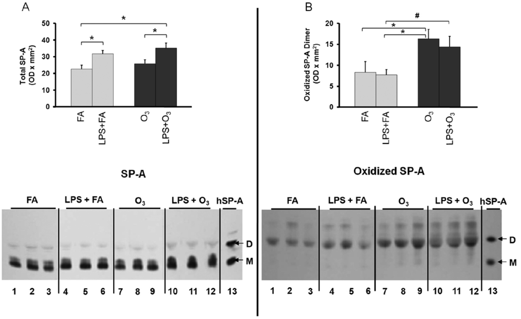
FIGURE 4.
The effects of lipopolysaccharide (LPS) with or without ozone (O3), O3 alone or filtered air (FA) alone on the level of surfactant protein A (SP-A) (Panel A) and oxidized SP-A (Panel B) in the bronchoalveolar lavage (BAL) of wild type (WT) mice. The mice received 2 ng of LPS intrapharyngeally in 50 µl saline, then 13 h later they were exposed to either FA (gray bars) or O3 (2 ppm for 3 h) (black bars) and subjected to BAL 4 h after ozone exposure or 20 h after LPS treatment. SDS gel electrophoresis was performed on concentrated samples from 200 µl of BAL fluid and the proteins transferred to membranes as described in Methods. Levels of SP-A (Panel A; top) and oxidized SP-A (Panel B; top) were measured by densitometry of Western blots. For each condition n=6 except for FA, where n=5. At the bottom of panels A and B a representative immunoblot (Panel A) and an oxyblot (Panel B) are shown. A reference lane is shown that contains immunostained human alveolar proteinosis SP-A (hSP-A) with bands of monomeric (M) and dimeric (D) SP-A marked in each panel. The position of the oxidized SP-A dimer bands was determined by comparison with an identical immunoblot stained with an antiserum to SP-A. The values depicted are the mean ± S.D. Significant (p≤0.05) differences between LPS + FA and LPS + O3 treated mice are indicated by the pound sign (#) and other comparisons by an asterisk (*).

