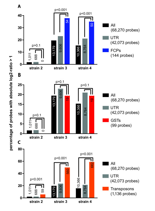
Figure 6.
Percentage of variable probes (i.e. probes exhibiting an absolute log2-ratio of test to reference strain > 1) belonging to different groups: (A, blue) fucoxanthin chlorophyll a/c binding proteins (FCPs), (B, red) glutathione S-transferases (GSTs), and (C, orange) TEs (Transposons). The higher the bar, the higher the degree of variability in a particular strain or group of probes. As a comparison, each graph shows also the percentage of variable probes among all probes (black) and only UTR probes (grey). P-values were calculated using a binomial test in comparison to both all probes and only UTR probes.

