Figure 4.
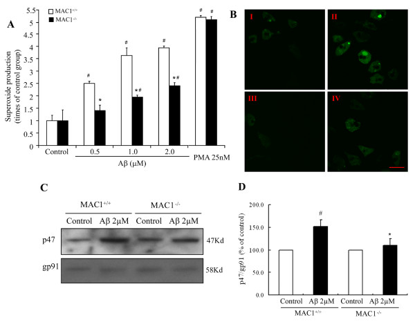
MAC1 mediates activation of PHOX and production of superoxide. (A) Microglia-enriched cultures from MAC1+/+ and MAC1-/- mice were treated with vehicle medium (control group), Aβ or PMA for 10 min. Extracellular superoxide generation was measured by the SOD-inhibitable reduction of tetrazolium salt, WST-1. (B) Microglia-enriched cultures from MAC1+/+ and MAC1-/- mice were incubated with HBSS containing 5 μM DCFDA in the dark for 1 hour. Then cells were treated with vehicle medium (control group) or Aβ for 10 min. Fluorescent images were captured using Zeiss 510 laser scanning confocal microscope. Scale bar: 25 μm. (C) Western blot assays for p47phox levels in membrane fractions of microglia from MAC1+/+ and MAC1-/- mice 10 min after vehicle medium (control group) or Aβ treatment. (D) Densitometry analysis was performed with values of p47phox normalized to loading control and further normalized to control levels. Data are presented as mean ± SEM from three independent experiments. #: p < 0.05 relative to corresponding vehicle-treated control cultures. *: p < 0.05 relative to MAC1+/+ cultures after same treatments.

