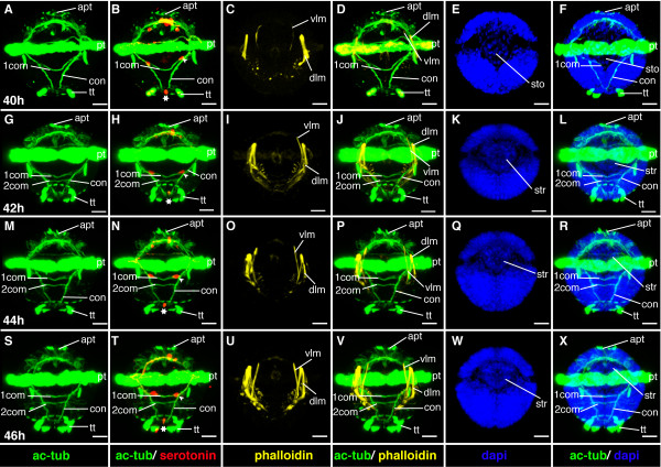
Figure 13.
Ventral nerve cord and muscle development of P. dumerilii, 40-46 hpf, ventral view, anterior up. The age of the larvae in each row is given in the lower left corner of the first picture of each row. The displayed staining is indicated at the bottom of each column. A, G, M, S: The second commissure (2com) of the ventral nerve cord is formed at 42 hpf. B, H, N, T: In addition to the unpaired serotonergic cell at the posterior end of the larva (white star), a pair of serotonergic neurons becomes visible at the first commissure (white arrow head). C, D, I, J, O, P, U, V: The dorsal and ventral longitudinal muscles (dlm and vlm) increase rapidly in length. E, F, K, L, Q, R, W, X: The stomodeal rosette (str) is formed and gets into a more anterior position. CLSM microscopy, maximum projection, Imaris surpass mode. Scale bar in all images 20 μm. Further abbreviations see abbreviations list.

