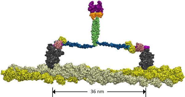
Figure 6.
Present model of the dimerized full-length myosin VI. The color scheme is adopted from Figure 1, except for the motor domain, which is shown in dark grey. MT domain (green) is the key dimerization region; the unfolded three-helix PT domain (blue) provides myosin VI with sufficient step length for the two motor domains (dark grey) to span a 36 nm step distance on the actin filament. There is a slight (~10 Å) offset in the relative placement of the two MT domain segments.

