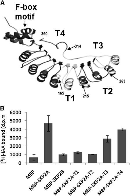
Figure 4.
Identification of the Auxin Binding Region in the SKP2A Structure.
(A) An orthogonal view of a ribbon diagram of the modeled structure of SKP2A using the structure of human Skp2. Arrows indicate the different deletions generated for auxin binding analyses (T4, T3, T2, and T1), and the last amino acid of the truncated proteins is indicated by a number.
(B) These truncated versions (MBP-SKP2A-T1, T2, T3, and T4), MBP, MBP-SKP2A, and MBP-SKP2B were expressed in bacteria and then incubated in the presence of 50 nM [3H]-IAA. The retained [3H]-IAA in the amylose beads after three washes was measured by scintillation counting. Each value is the mean of three independent measures, and the error bars correspond to the sd. d.p.m., disintegrations per minute.

