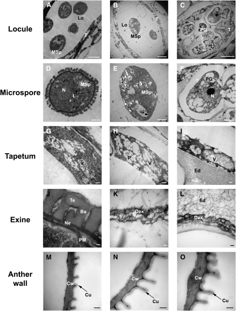
Figure 9.
Transmission Electron Micrographs of Wild-Type (Col-0) and pksa-1 pksb-3 Anthers and Pollen.
(A), (D), (G), (J), and (M) Microspore structure, tapetum structure, exine formation, and outer wall of anther epidermis at anther stage 9 in Col-0 wild-type plants.
(B), (E), (H), (K), and (N) Microspore structure, tapetum structure, exine formation, and outer wall of anther epidermis at anther stage 9 in pksa-1 pksb-3 plants.
(C), (F), (I), (L), and (O) Pollen grain structure, tapetum structure, exine formation, and outer wall of anther epidermis at anther stage 11 in pksa-1 pksb-3 plants.
Ba, bacula; Cu, cuticle; Cw, cell wall; Dex, defective exine structure; Ed, electron-dense material; Ex, exine; L, lipid body; Lo, locule; MSp, microspore; N, nucleus; Ne, nexine; P, plastid filled with plastoglobuli; PG, pollen grain; PM, plasma membrane; T, tapetal cell; Te, tectum; V, vacuole containing electron-dense material. Bars = 10 μm in (A) to (C), 2 μm in (D) to (F), 500 nm in (G) to (I) and (M) to (O), and 100 nm in (J) to (L).

