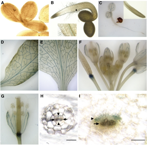
Figure 5.
Expression Pattern of NaKR1pro:NaKR1-GUS Specifically in the Phloem Region of the Vasculature.
(A) No GUS expression was detected in imbibed seeds prior to germination. Seed coat was removed to facilitate observation.
(B) GUS staining was visible in the vasculature of a 1-DAG seedling in the region of root-hypocotyl boundary (indicated by arrow and shown at higher magnification in the inset).
(C) A 2-DAG seedling. Inset shows root tip at higher magnification.
(D) Mature rosette leaf.
(E) Cauline leaf.
(F) Floral tissue. Note that the strongest staining is in the pedicel of developing siliques and in the sepals.
(G) Sepals and stamen filaments in an opening flower.
(H) Cross section of a young root after GUS staining. The positions of the phloem and xylem region are indicated (by the arrowheads and arrows, respectively). Note that GUS expression was found in the phloem cells.
(I) Cross section of a rosette leaf petiole after GUS staining. The position of the phloem and the xylem regions of the main vasculature are indicated as in (H). Note that GUS expression was limited to the phloem tissue. Bars in (H) and (I) = 50 μm.

