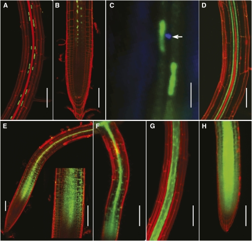
Figure 6.
NaKR1 Is Expressed in Companion Cells, and NaKR1-GFP Is Phloem Mobile.
Confocal images (except in [C]) of Arabidopsis tissue stained briefly with propidium iodide.
(A) NaKR1pro:histone2B-GFP localization in companion cell nuclei of Col-0 primary root.
(B) NaKR1pro:histone2B-GFP localization in the proximal meristem region of the primary root meristem.
(C) Epifluorescence image of NaKR1pro:histone2B-GFP expression in Col-0 root. Aniline blue staining shows sieve plate (indicated by arrow) of the adjacent sieve element.
(D) NaKR1pro:NaKR1-GFP expression in a mature, complemented nakr1-1 root.
(E) NaKR1pro:NaKR1-GFP fluorescence in the proximal meristem region of a primary root of complemented nakr1-1. Inset is magnified meristem region.
(F) NaKR1pro:NaKR1-GFP expression in the lateral root of complemented nakr1-1.
(G) Free GFP expressed using the SUC2 promoter showing fluorescence in the mature primary root of Col-0.
(H) Expression of free GFP under control of the SUC2 promoter in a primary root meristem of Col-0. Scale bars indicate 100 μm except for C in which the bar is 10 μm.

