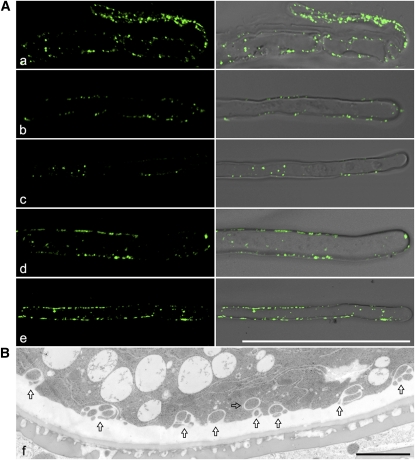
Figure 15.
Immunofluorescent and Structural EM Detection of EXPO in Other Cell Types.
(A) Shown are examples of EXPO detected by anti-Exo70E2a antibodies in wild-type Arabidopsis root tip cells (a) and root hair cells (b to e). Bars = 50 μm.
(B) Structural EM detection of EXPO in an ultrathin section prepared from high-pressure freezing/frozen-substituted wild-type tobacco pollen grains. Arrows indicated examples of EXPO. Bars = 2 μm.
[See online article for color version of this figure.]

