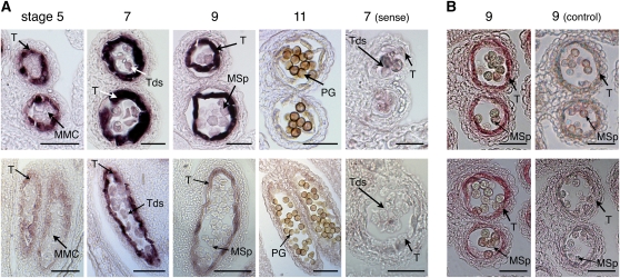
Figure 2.
Tapetum-Specific Expression of TKPR1 and TKPR2.
Top panels: TKPR1 expression; bottom panels: TKPR2 expression.
(A) TKPR1 and TKPR2 mRNAs were localized by in situ hybridization of gene-specific antisense probes to sections of wild-type (Col-0) flowers. Sense probes were used for controls. Stages of anther development are according to Sanders et al. (1999). Dark precipitates indicate hybridization of the probe. Stage 7 shows highest hybridization signals for both of TKPR1 and TKPR2 in the tapetum, but TKPR2 expression was more restricted temporally.
(B) Specific antibodies detected TKPR1 and TKPR2 protein accumulation in the tapetum of anthers at stage 9 of development. Controls were performed using preimmune sera.
MMC, microspore mother cells; Tds, tetrads; T, tapetum; MSp, microspores; PG, pollen grain. Bars = 70 μm.

