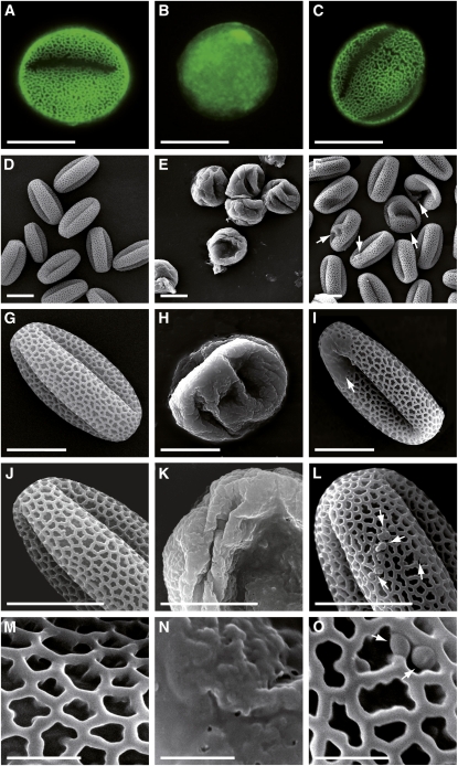
Figure 6.
Comparison of Exine Architecture in Wild-Type, tkpr1-2, and tkpr2-1 Pollen.
(A), (D), (G), (J), and (M) Wild-type pollen.
(B), (E), (H), (K), and (N) tkpr1-2 pollen.
(C), (F), (I), (L), and (O) tkpr2-1 pollen; arrows indicate exine defects.
(A) to (C) Epifluorescence microscope images of wild-type and mutant pollen. Pollen was stained with the fluorescent dye auramine O and visualized using fluorescein isothiocyanate settings.
(D) to (O) Scanning electron micrographs of wild-type and mutant pollen grains.
Bars = 10 μm in (A) to (L) and 2 μm in (M) to (O).
[See online article for color version of this figure.]

