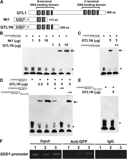
Figure 6.
N-Terminal DNA Binding Domain of GTL1 Binds to PHYA and SDD1 Promoters.
(A) Schematic diagram of the GTL1 protein, showing the four helical, N- and C-terminal DNA binding domains (represented as gray cylinders). Two protein fragments (Nt1 and GTL1N) were fused with MBP.
(B) Nt1 and GTL1N polypeptides and a biotin-labeled rice PHYA promoter fragment (400 ng) were used for EMSA. In (B) to (E), free probe and GTL1N-probe complexes are indicated by an asterisk and arrows, respectively.
(C) Unlabeled PHYA promoter (2 μg) was used as a competitor to determine the specificity of DNA binding activity for GTL1N.
(D) GTL1N polypeptide was used for EMSA with a biotin-labeled SDD1 promoter fragment (250 ng). Unlabeled probes (250 ng [+] and 1 μg [++]) were used as competitors. An unlabeled mutant version of the SDD1 promoter (GG → CC) was used as a noncompetitor.
(E) The mutant version of the SDD1 promoter (GG → CC) was labeled with biotin (250 ng) and used for EMSA with GTL1N polypeptides.
(F) ChIP analysis was conducted to determine the in vivo interaction between GTL1 with the SDD1 promoter. Input is chromatin before immunoprecipitation. Anti-GFP antibody was used to precipitate chromatin associated with GTL1-GFP. Mouse IgG was used as a negative control for the specificity of immunoprecipitation. The SDD1 promoter region associated with GTL1 was amplified by PCR using SDD1 promoter-specific primers for three different regions (1, 2, and 3). Region 1 (−1136 to −1400) is distal to the GT3 box. Region 2 (−538 to −824) is adjacent to, but does not contain, the GT3 box. Region 3 (−279 to −556) includes the GT3 box in the SDD1 promoter.

