
Figure 2. Treatment with NVP-BEZ235 inhibits tumor growth in FL mouse xenograft model by targeting the PI3K-Akt-mTOR-S6K pathway.
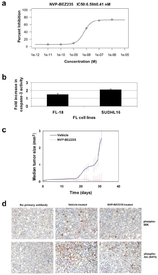
a. FL-18 cells were treated with various concentrations of NVP-BEZ235 for 72 hours. The IC50 for FL-18 growth inhibition was calculated at 6.59nM. b. FL-18 and SUDL16 cells were treated with either DMSO or 25 nM NVP-BEZ235 for 24 hours after which cells were harvested and subjected to a caspase-3 activation assay (Promega). The increase in caspase-3 activation in the NVP-BEZ235-treated cells compared to cells incubated with vehicle control is shown on the Y-axis. c. Treatment of FL-18 tumors with NVP-BEZ235 suppressed tumor growth in vivo. Tumor volume is depicted on the y-axis and time in days is shown on the x-axis. All animal procedures were performed in accordance with the UNC-CH IACUC. Briefly, NOD.CB17-Prkdcscid/J or 4-5-week-old athymic nude-Foxn1ˆnu female mice were injected with anti-asialo GM1 antibody (Wako) intraperitoneally. Twenty four hours later, the mice were subcutaneously injected 1×10⁷ or 1×10⁶ FL-18 cells per mouse. Treatment with NVP-BEZ235 or vehicle was started upon the development of palpable tumors. NVP-BEZ235 was dissolved in a 1:9 v/v mixture of 1-methyl-2-pyrrolidone (Fluka) and polyethylene glycol 300 (Sigma). A dose of 45 mg/kg or equal volume of the vehicle was administered orally 5-6 days per week as previously described (11). A total of eighteen mice were treated with NVP-BEZ235, and seventeen mice were treated with vehicle. Tumor volume (LxWxD) was calculated daily and animals were sacrificed when the tumor volumes of control mice reached the maximum allowed by IACUC. Tumors were excised from mice, fixed and sectioned. d. Immunohistochemistry of tumors from vehicle- and NVP-BEZ235-treated mice. Tumors were fixed, sectioned, and stained with the antibodies against phospho-S6K and phospho-Akt (S473) described previously (10). Images are shown at 400X magnification. Sections stained without primary antibody served as a negative control.