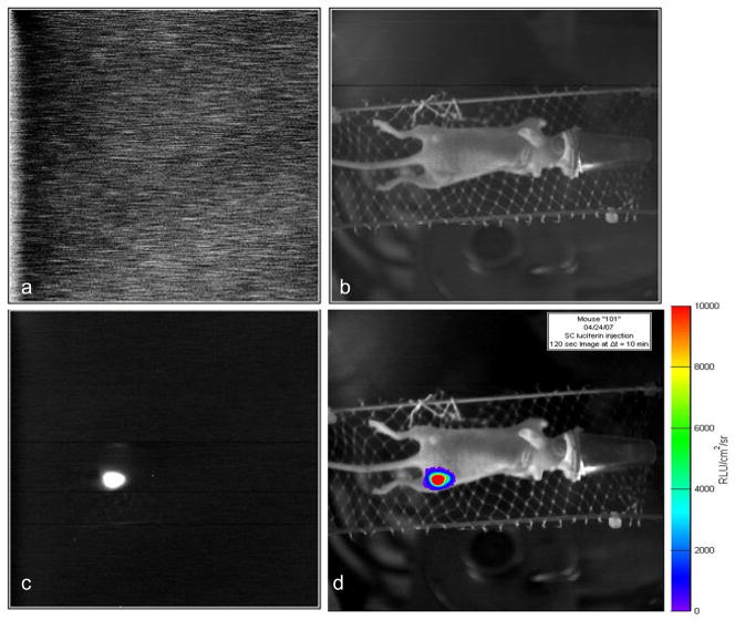
Figure 1. Images associated with BLI.
Images acquired with home-built Cyclops BLI system. A) Dark image to allow subtraction of background noise; b) Light image based on surface external illumination for anatomical registration; c) Bioluminescent image; c) Overlay of bioluminescent signal intensity on anatomical image.

