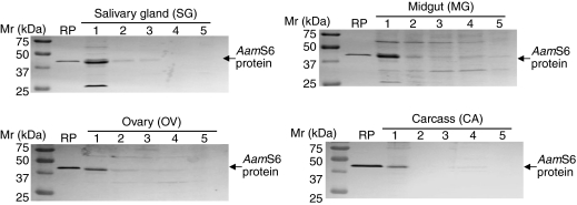
Fig. 4.
Spatial and temporal western blot analyses of native AamS6 protein in dissected tick organs. Total protein extracts of salivary gland, midgut, ovary and carcass (tick remnant after removal of SG, MG and OV) pooled from eight ticks per time point were subjected to western blot analyses using antibodies to rAamS6. Arrows denotes the position of the native AamS6 protein. Please note that membranes probed with pre-immune sera are not shown. Please refer to Fig. 3.

