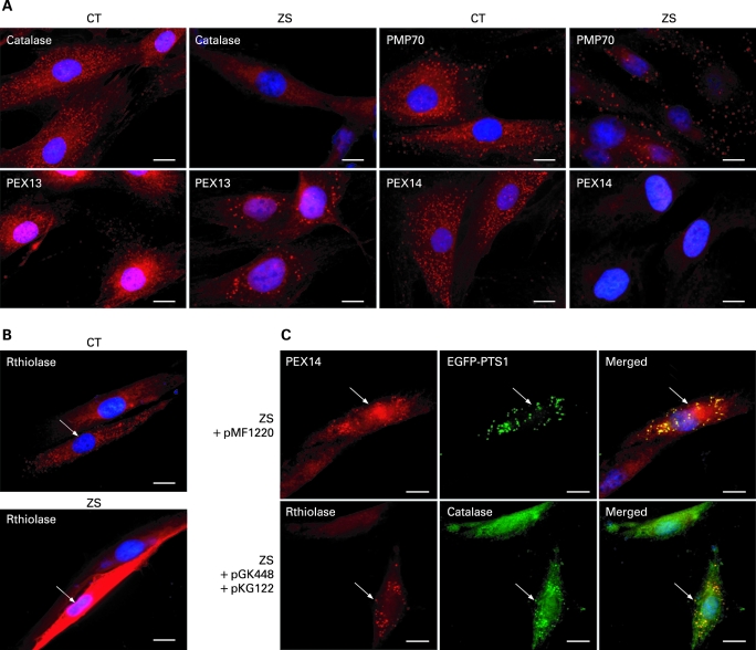
Figure 2. Immunocytochemical localisation of peroxisomal proteins in patient and control fibroblasts.
(A) Control (CT) and Zellweger patient (ZS) fibroblasts were processed for immunostaining with antibodies specific for catalase, PEX13, PMP70, or PEX14 (red). (B) As we experienced difficulty detecting endogenous thiolase in human skin fibroblasts, the cells were transfected with a plasmid coding for rat thiolase B (rthiolase) 2 days before immunostaining with anti-thiolase B antibodies. (C) Cultured patient fibroblasts were (co)transfected with pMF1220, a bicistronic plasmid encoding non-tagged human PEX14 and EGFP-PTS1, or pGK448 and pKG122, two monocistronic plasmids encoding non-tagged human PEX14 and rat thiolase B, respectively; after 2 days, the cells were processed for fluorescence microscopy using rabbit anti-PEX14 antibodies (upper row) or rabbit anti-thiolase B (red) and mouse anti-catalase (green) antibodies (lower row); note that catalase and EGFP-PTS1, two PTS1-containing proteins, as well as thiolase B, a PTS2-containing protein, display a punctate staining pattern indicating the reconstitution of functional peroxisomes. The nuclei were counterstained with DAPI (blue), and transfected cells are marked by arrows. Scale bar: 20 μm.

