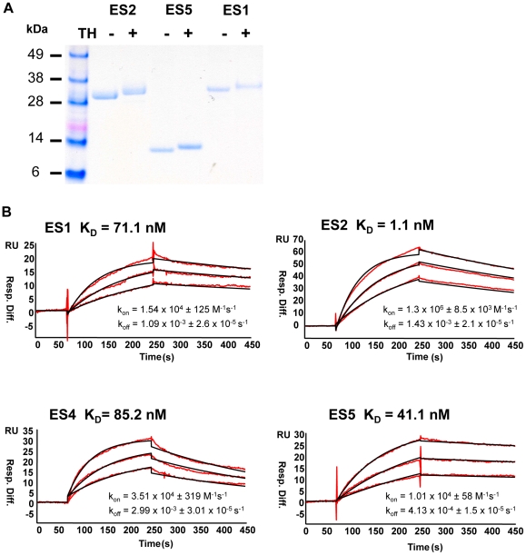
Figure 2. Biophysical characterization of the ES proteins.
(A) SDS-PAGE gel of ES proteins used as immunogens after affinity purification. The ES proteins possessing the C-terminal heterologous T cell helper residues are denoted “+TH”. (B) The recognition of the ES proteins by the 2F5 monoclonal antibody was assessed by surface plasmon resonance (SPR) in a Biacore 3000 instrument. In red, the observed data obtained by flowing the ES proteins as analytes over a CM5 chip to which the 2F5 IgG antibody was immobilized. In black, fit curves when a 1∶1 Langmuir model is applied to the observed data. Affinity constant values are indicated above the curves and the rate constants are denoted below the curves.

