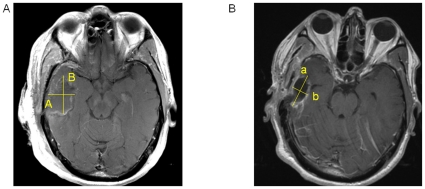
Figure 3. Detection of Enhancing Tumor Volume Despite Resection Cavity Collapse.
A) T1-weighted post-contrast axial image showing a resection cavity with rim enhancement. RECIST measurement would be A and Macdonald measurement would be “A * B”. B) T1-weighted post-contrast axial image showing the same patient 3 months postoperatively who had collapse of his resection cavity. RECIST measurement would be “a” and Macdonald measurement would be “a * b”, both of which would be smaller than the measurements from the initial scan above, but this change would be describing only the resection cavity configuration and not the underlying tumor burden.

