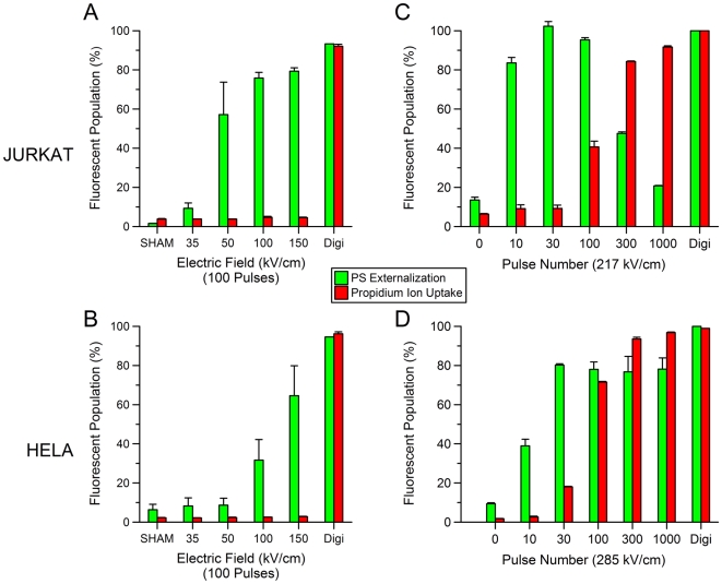
Figure 5. The effect of the E-field and pulse number on USEP-induced externalization of phosphatidylserine (PS) and uptake of propidium ions (Pr).
PS and Pr fluorescent expression for Jurkat (A,C) and HeLa (B,D) exposed to increasing electric fields between 0–150 kV/cm at 100 pulses per exposure and 0.005% digitonin. Externalization of PS appears in Jurkat at lower field amplitudes than in HeLa. Both cell lines exposed to digitonin stained positive for both PS and Pr. C and D show percent of Pr positive Jurkat and HeLa after exposure to increasing number of pulses at 217 and 285 kV/cm, respectively. (mean +/− s.d., n = 3 measurements of 25,000 cells).

