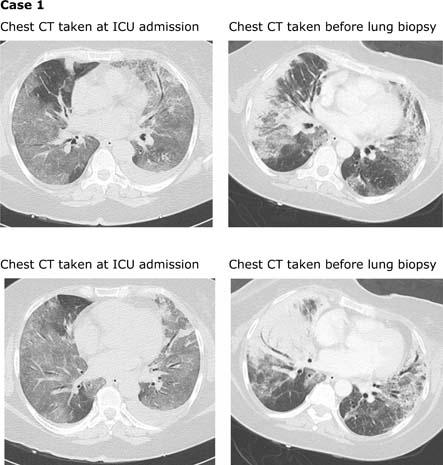
Figure 1.

Chest CT taken at ICU admission show diffuse ground glass opacities with some peripheral and peribronchovascular lobular consolidation. Chest CT taken before lung biopsy show foci of ground glass have evolved to bilateral consolidations, predominantly in a subpleural peripheral distribution. Areas of ground glass opacities have appeared in both upper lobes (not shown in these pictures). In left lower lobe, ground glass opacities has been replaced by fine reticular intralobular opacities with traction bronchioloectasis (suggesting initial fibrosis).
