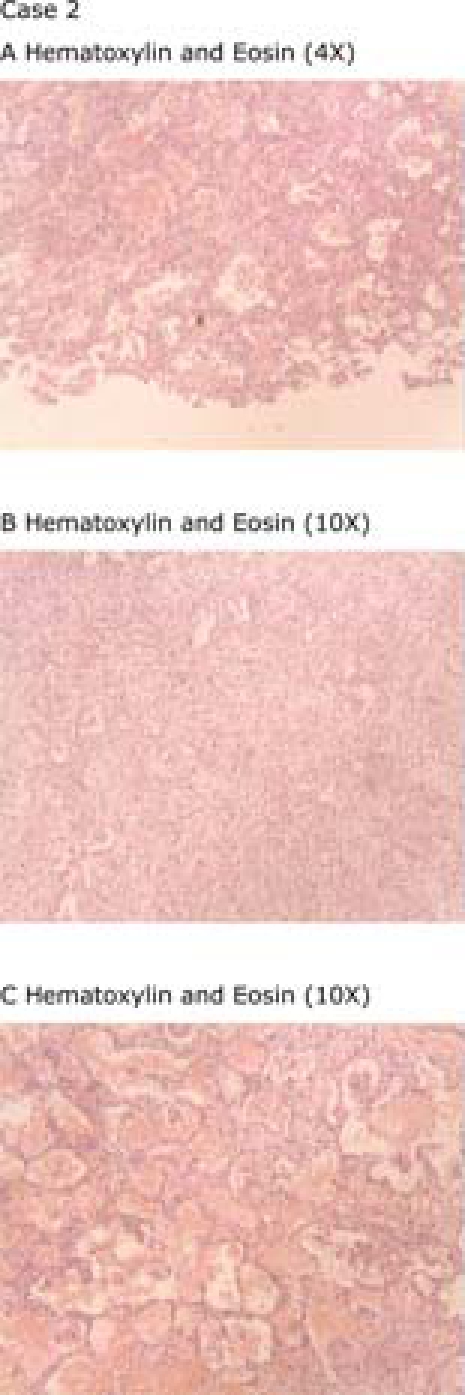
Figure 4.

Histopathological findings in Case 2.(A) Mild interstitial inflammatory lymphoplasmacytic cell infiltrate and sparse fibrin accumulation intraalveolar. (B) Solid area of prominent organizing pneumonia and athelectasia. (C) Intraalveolar oedema and haemorrhage admixed with macrophages and Masson bodies.
