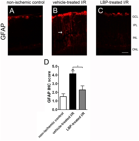
Figure 4. Lower level of glial fibrillary acidic protein (GFAP) activation in the LBP-treated I/R retina.
(A–C) Immunohistochemistry of GFAP. I/R induced intense GFAP immunoreactivity in Muller cell processes of the vehicle-treated I/R retina (arrows in B), but not of the LBP-treated I/R retina (C). Results were confirmed by semi-quantification of IHC in (D). ##p<0.01 vs. non-ischemic control retina; *P<0.05 vs. vehicle-treated I/R retina. Scale bar, 25 µm.

