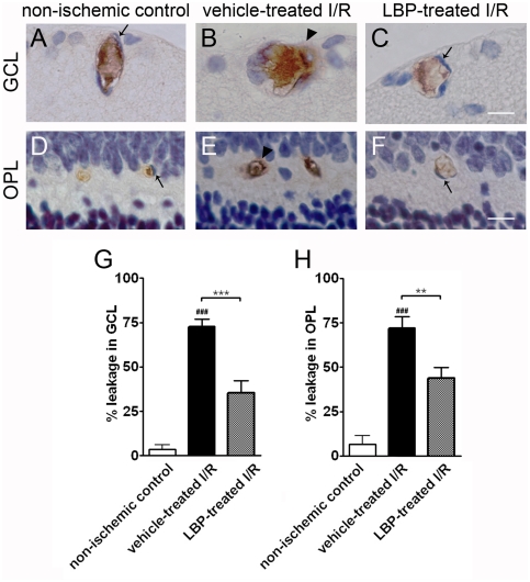
Figure 6. The number of leaky blood vessels in the LBP-treated I/R retina was comparable with that in the non-ischemic control retina.
(A–C) IgG extravasations in blood vessels in GCL. (D–F) IgG extravasations in blood vessels in OPL. In non-ischemic control retina, IgG staining was confined inside the blood vessel lumen demarcated by endothelial cells (arrows in A & D). However, I/R induced blood vessel leakage in both GCL and OPL (arrow heads in B & E) in the vehicle-treated I/R retina but not in the LBP-treated I/R retina (arrows in C & F). (G, H) Quantification of blood vessel leakage in GCL (G) and OPL (H). More leaky blood vessels were observed in GCL (G) and OPL (H) of the vehicle-treated I/R retina. However, LBP treatment decreased the I/R-induced blood vessel damage, evident from fewer leaky blood vessels. ###p<0.001 vs. non-ischemic control retina; **p<0.01, ***p<0.001 vs. vehicle-treated I/R retina. Scale bar, 25 µm.

