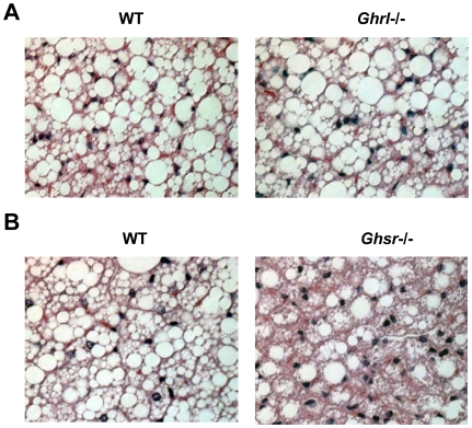
Figure 4. Morphology of interscapular brown adipose tissue (BAT) of older WT, Ghrl-/-, and Ghsr-/- mice.
(A): The morphology was very similar between older Ghrl-/- and their WT counterparts. (B): BAT of older Ghsr-/- mice showed higher percentages of multilobular adipocytes and increased cellularity (dark blue nuclei). These are representative H & E staining of BAT paraffin sections from 4–8 mice.

