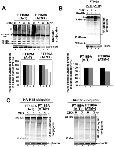
Figure 1. Protein turnover is reduced in A-T cells.
A. FT169A (A-T) (lanes 1–4) and FT169A (ATM+) (lanes 5-8) cells were treated with the protein synthesis inhibitor CHX (10 µg/ml) for 0, 1, 3, and 6 hours. Cell lysates were analyzed using discontinuous (5%/15%) SDS-PAGE followed by immunoblotting with anti-ubiquitin antibody. The symbols * and ** mark the position of high-molecular-weight (HMW) polyubiquitylated proteins. Quantitation of the high-molecular-weight (HMW) polyubiquitylated proteins (shown as **) is shown in the bar graph. B. FT169A (A-T) (lanes 1 and 2) and FT169A (ATM+) (lanes 3 and 4) cells were transfected with HA-ubiquitin as described in Methods. Forty-eight hours post-transfection, cells were treated with the protein synthesis inhibitor CHX (marked on top of each lane) for 6 hours. Cell lysates were analyzed using 15% SDS-PAGE followed by immunoblotting with anti-HA antibody. The symbol ** marks the position of polyubiquitylated proteins (compressed due to the gel electrophoresis conditions). Quantitation of the high-molecular-weight (HMW) polyubiquitylated proteins (shown as **) is shown in the bar graph. C. FT169A (A-T) and FT169A (ATM+) cells were transfected with HA-Lys48-only (left panel) and Lys63-only (right panel) ubiquitin constructs. Thirty hours post-transfection, cells were treated with the protein synthesis inhibitor CHX (marked on the top of each lane) for 3 hours and then analyzed by immunoblotting with anti-HA antibodies as described above. All the experiments were repeated at least three times and the representative experiments are shown.

