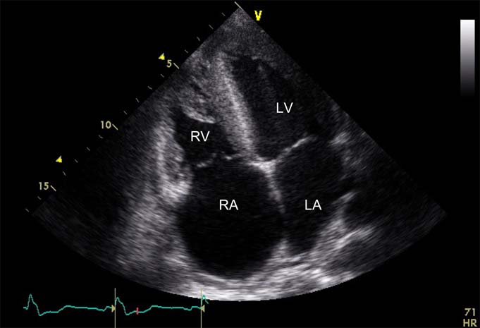
Figure 1.

Transthoracic echocardiography. Image from the apical four-chamber view illustrating marked hypertrophy of the left ventricle (LV) and the right ventricle (RV), hyperechogenic myocardium, RV dilatation as well as dilatation of the right atrium (RA) and, to a lesser extent, the left atrium (LA).
