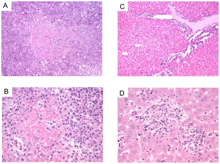
Figure 2. Histopathology of spleen and liver from AHSV-4 infected IFNAR −/− mice.
Liver and Spleen. Hematoxylin-eosin staining. Representative examples of the microscopic lesions found on liver and spleen of AHSV-4 infected IFNAR −/− mice are shown. Spleen and liver samples were collected from IFNAR −/− mice, on day 7 post-infection with AHSV-4. The spleen shows loss or its normal structure (a) and abundant deposits of amyloid (a,b). The liver presents a mononuclear inflammatory infiltrate in the portal triad (c) and foci of necrosis (d).

