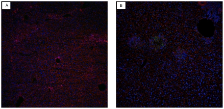
Figure 5. Detection of AHSV- antigen in liver tissues of AHSV-4 infected IFNAR −/− mice.
Livers of normal uninfected (a) and AHSV-4 infected (b) mice were processed for immuno-fluorescence as indicated in Materials and Methods. The presence of AHSV antigen in the liver (green fluorescence) accumulates in foci of cells that resemble the foci of necrosis observed in hematoxylin-eosin sections.

