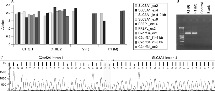
Figure 1. Genetic analysis of the atypical hypotonia–cystinuria syndrome (HCS) patients.
(A) Quantitative polymerase chain reaction (PCR) analysis on genomic DNA from both siblings and control samples. Only relevant amplicons are shown. (B) Junction fragment PCR spanning the breakpoint. P1 (M), patient 1 (male); P2 (F), patient 2 (female). (C) The sequence at the joining of the deletion ends. The junction fragment PCR product derived from patient DNA was subjected to sequencing with the primer located in C2orf34 intron 1 used for the generation of the junction fragment. The two bases CA could be derived from either side, as they are present in both.

