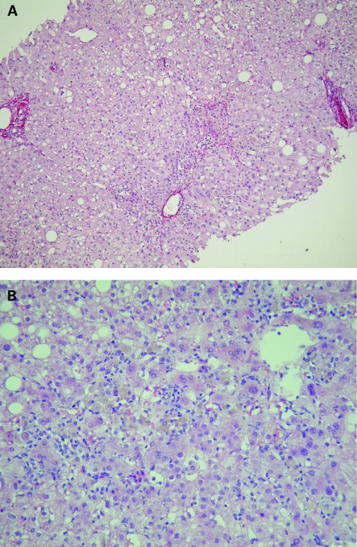
Figure 2.
Liver lesions. (A) Sirius red stain. Mild sinusoidal pericentrolobular fibrosis (black pointer) and normal portal tract (arrows). (B) Haematoxylin and eosin stain. Centrilobular damages: spotty (arrow) and confluent (black pointer) necrosis with lymphocytes, scattered eosinophils and macrophages with pigments.

