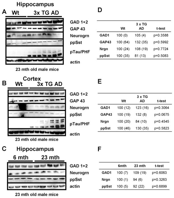
Fig. 7.
Levels of proteins involved in inhibitory neurotransmission are unchanged in the cortex and hippocampus of a mouse model of Alzheimer’s disease. (A and B) Levels of GAD1, GAP43, neurogranin (nrgn), and preprosomatostatin (pp Sst) did not differ in the hippocampus (A) or cortex (B) between 23-month-old male wild type and 3xTgAD mice. Levels of phosphorylated tau protein were elevated in the 3xTgAD mice, indicating their advanced stage of disease. (C) Levels of GAD1, GAP43, neurogranin, and preprosomatostatin in the hippocampus did not differ between 6-month-old and 23-month-old wild type mice. (D, E, and F) Densitometric quantification of immunoblots. Average densitometric intensity (+ SEM) displayed as percent of wild type animals (D and E) or 6-month-old animals (D) with corresponding p values for t-test. Bold p values indicate statistical significant differences.

