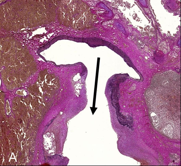
Figure 3.

Elastica-van Giesson-staining (1×). This special staining to detect collagen (red) and elastic fibres (black) highlights vascular architecture with extravascular erythrocytes in the surrounding lung parenchyma (brown). The arrow base is located within the normal artery part while the arrow head marks the ectatic vessel wall with degradation of fibres and smooth muscle cells by granulocytes indicating mycotic genesis of the aneurysm.
