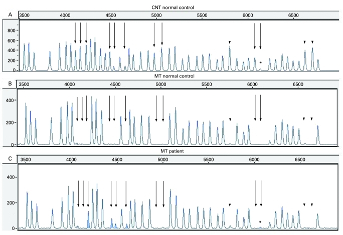
Figure 1.
The methylation-specific multiplex ligation-dependent probe amplification (MS-MLDPA) profiles. (A) Copy number test (CNT) in a normal male control. Straight arrows: methylation-specific FMR1 probes; dotted arrows: methylation-specific FMR2 probes; arrowheads: digestion controls. The fragment marked by asterisk is FMR1 exon 1 specific probe, which gives low signal and might be less reliable (manufacturer’s communication). (B) Methylation test in a normal control. All methylation-specific bands (pointed by different arrows) are missing as a result of HhaI digestion. (C) Methylation test (MT) in the presented patient. The five FMR1 exon 1 methylation-specific probes gave a signal (peaks are given in grey and pointed by straight arrows), as a result of hypermethylated full mutation, not digested by HhaI. The remaining methylation-specific fragments (FMR2 and digestion controls) are missing showing that the digestion was successful and there is no hypermethylation along the FMR2 gene.

