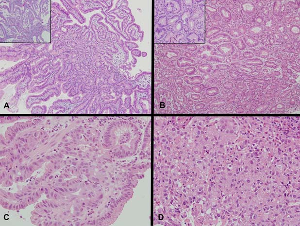
Figure 1.

(A) Sections demonstrating a polypoid lesion arising among distorted duodenal villi composed of tubular glands lined by columnar epithelium with basal nuclei and pale to oeosinophilic ground glass cytoplasm (H&E ×100, inset PAS with Diastase, ×200). No apical mucin cap is demonstrated within the epithelium of the glands (inset). (B) Sections demonstrating abrupt transition of typical pyloric type glands to glands with low-grade dysplasia showing irregularity of gland outline with elongated, pseudostratified and mildly hyperchromatic nuclei (H&E ×100, inset H&E ×400). Higher magnification showing two glands in the centre with abrupt transition within the same gland from typical bland cytology of pyloric type epithelium to low-grade dysplasia with mildly hyperchromatic and pseudostratified nuclei (inset). (C) High-grade dysplasia with irregular papillary structures and fused irregular glands lined by epithelium showing enlarged nuclei with loss of polarity, nuclear pleomorphism and enlarged nucleoli (H&E ×400). Increasing numbers of mitotic figures are seen. (D) Intramucosal carcinoma with syncytial growth of back-to-back and fused poorly formed microglands and solid clusters of malignant cells with increasing nuclear pleomorphism, loss of polarity and nucleolar prominence (H&E ×400).
