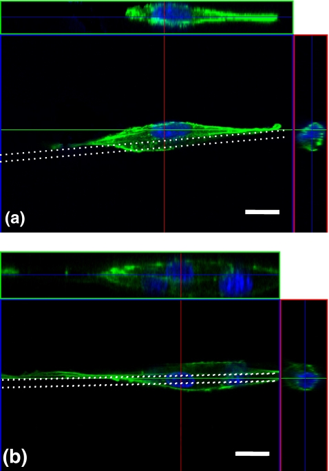
Fig. 4.
Confocal fluorescence microscope images of three-dimensional cell morphology and cross-sectional images of a single cell (a) and multiple cells (b). Stained actin cytoskeleton (Alexa 488-phalloidin: green) and nuclei (DAPI: blue). White dotted lines indicate fiber scaffolds of 8 μm in diameter. Scale bars: 20 μm

