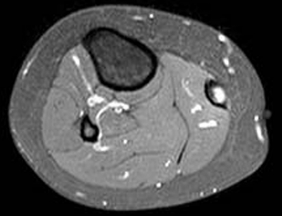
Figure 14.
T2 fat suppressed MRI images of the torn tendon to the medial head of gastrocnemius. The scans show progressive rounding and widening of the tendon distally due to a longitudinal split and tear mid-substance. The surrounding muscle (medial head of gastrocnemius) is normal with no tear, inflammation or scarring.

