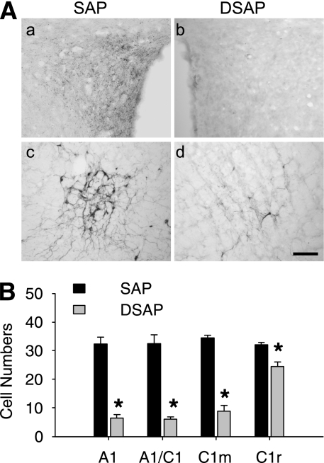
FIG. 4.
Lesion effect of DSAP on hindbrain catecholamine neurons. A: Representative IHC images of DBH staining in a SAP-injected (a and c) or DSAP-injected (b and d) rat in PVH (a and b) or the hindbrain A1/C1 region (c and d). Scale bar = 0.5 mm. B: DBH-positive cells in each of the hindbrain regions. *P < 0.01 vs. SAP rats (n = five rats in each group).

