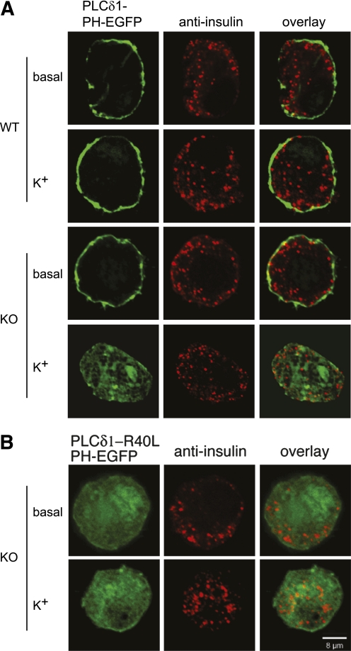
FIG. 3.
Plasma membrane PIP2 is not maintained in KO β-cells on depolarization. A: β-cells isolated from WT and KO islets were transfected with a plasmid expressing the PIP2 sensor PLCδ1-PH-EGFP, cultured for 2 days, depolarized with extracellular K+ for 5 min, fixed, and imaged using confocal microscopy. Pancreatic β-cells were identified using an anti-insulin antibody in combination with a red fluorescent secondary antibody. B: KO β-cells expressing a mutant PIP2 sensor (PLCδ1-PH-R40L-EGFP) that does not exhibit PIP2 binding activity were processed as in A. Images were taken through the midpoints of the cells and are representative of the typical transfected, insulin-positive cells observed in the experiments (n > 100). The experiment was repeated at least three times using two animals each time (n = six mice), and similar outcomes were obtained. Greater than 90% of the cells examined responded as shown in the images in each condition. (A high-quality color representation of this figure is available in the online issue.)

