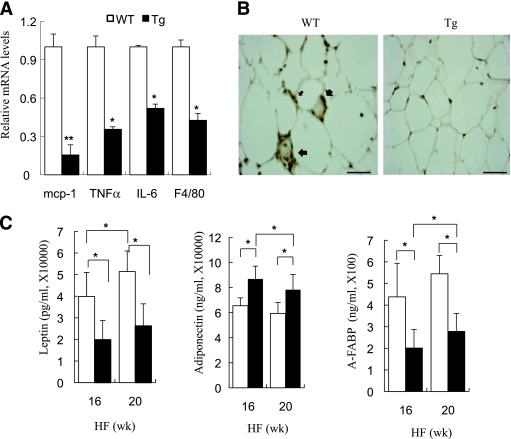
FIG. 7.
Adipose tissue–specific inactivation of JNK alleviated the HFD-induced inflammatory changes. A: Quantitative RT PCR analysis for the gene expression of F4/80 and the inflammatory cytokines MCP-1, TNF-α, and IL-6 in epididymal fat. Expression levels of all genes were normalized against 18S rRNA. B: Immunohistochemical staining for F4/80 in epididymal fat tissue sections from aP2-dn-JNK transgenic (Tg) mice and wild-type (WT) littermates. Scale bar: 50 μm. C: Serum leptin, adiponectin, and A-FABP levels in overnight-fasted aP2-dn-JNK transgenic mice and wild-type littermates were measured by an enzyme-linked immunosorbent assay after being fed an HFD for 16 and 20 weeks, respectively. □, wild-type littermates; ■, aP2-dn-JNK transgenic mice. *P < 0.05; **P < 0.01 vs. wild-type littermates (n = 6–8). (A high-quality color representation of this figure is available in the online issue.)

