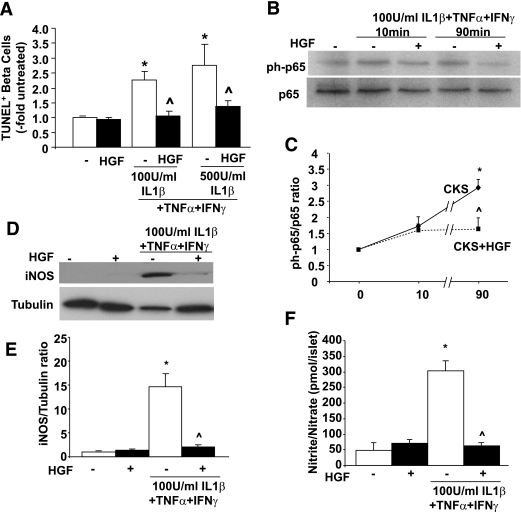
FIG. 7.
Protective effect of HGF on primary mouse β-cells treated with cytokines. A: Mouse islet cell cultures were treated with or without 25 ng/mL HGF and 100 or 500 units/mL IL-1β, 1,000 units/mL TNF-α, and 1,000 units/mL IFN-γ for 24 h. Results are means ± SE of five experiments in duplicate. B: Representative Western blot displaying phospho- and total p65 levels in protein extracts from mouse islets treated with or without 25 ng/mL HGF and 100 units/mL IL-1β, 1,000 units/mL TNF-α, and 1,000 units/mL IFN-γ for different time periods. C: Densitometric quantitation of phospho- and total p65 in four Western blots performed with four different protein extracts. D: Representative Western blot displaying iNOS and tubulin levels in protein extracts from mouse islets treated with or without the same doses of cytokines and HGF for 24 h. E: Densitometric quantitation of iNOS expression in three Western blots performed with three different protein extracts. F: Medium nitrite levels secreted from islets treated with or without the same doses of cytokines and HGF for 24 h. *P < 0.05 vs. untreated and ^P < 0.05 vs. cytokine-treated but HGF-untreated cells. CKS, cytokines.

