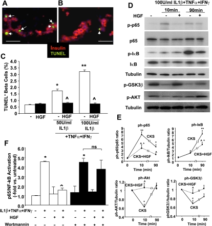
FIG. 8.
Protective effect of HGF on primary human β-cells treated with cytokines. A: Representative images of human islet cell cultures treated with 100 units/mL IL-1β, 1,000 units/mL TNF-α, and 1,000 units/mL IFN-γ for 24 h in the absence or (B) presence of 25 ng/mL HGF and stained for TUNEL (green), insulin (red), and DAPI (blue). Arrows indicate TUNEL-positive β-cell nuclei. Scale bar = 25 μm. C: Quantitation of TUNEL-positive β-cell nuclei in five experiments per duplicate performed with human islet cell cultures from five different donors treated with 50 or 100 units/mL IL-1β, 1,000 units/mL TNF-α, and 1,000 units/mL IFN-γ for 24 h. D: Representative Western blots displaying the expression of phospho- and total p65, phospho- and total IκB, phospho-GSK-3β, phospho-AKT, and tubulin in protein extracts from human islets treated with or without HGF and 100 units/mL IL-1β, 1,000 units/mL TNF-α, and 1,000 units/mL IFN-γ. E: Densitometric quantitation of these proteins in four Western blots performed with four different human islet extract samples per time point obtained from four different donors. F: Activation of p65/NF-κB in human islet extracts treated with 50 units/mL IL-1β, 1,000 units/mL TNF-α, and 1,000 units/mL IFN-γ for 10 min and with or without 25 ng/mL HGF and assessed by an ELISA-based TransAM assay measuring p65/NF-κB binding activity (see research design and methods). In some cases, human islets were pretreated for 30 min with 10 nM Wortmannin. Results are means ± SEM of three experiments in triplicate. *P < 0.05 and **P < 0.01 vs. untreated and ^P < 0.05 vs. cytokine treated. CKS, cytokines; ns, not significant. (A high-quality digital representation of this figure is available in the online issue.)

