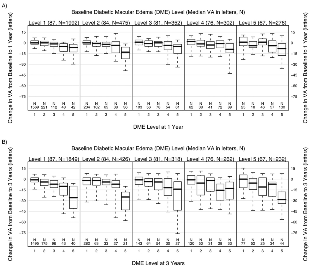
FIGURE 1.
Change in median VA (number of letters) between baseline and 1 year (1A) and between baseline and 3 years (1B) in eyes assigned to deferral of photocoagulation, by DME severity level on the five-step scale (see Table 1) at baseline and at follow-up. Boxes show the 25th and 75th percentiles, whiskers the 10th and 90th percentiles, and the line within box the median.

