Fig. 17.

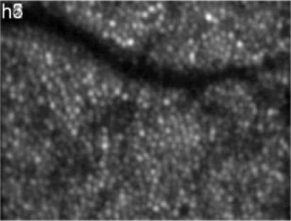
En-face image of the RPE, averaged over the measurement period of 7 hours (depth integrated 12µm around the peak of the RPE position) OCT images. (Image extension: ~0.94°x0.7°, retinal eccentricity: ~4° nasal from the fovea)

En-face image of the RPE, averaged over the measurement period of 7 hours (depth integrated 12µm around the peak of the RPE position) OCT images. (Image extension: ~0.94°x0.7°, retinal eccentricity: ~4° nasal from the fovea)