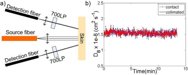
Fig. 2.

a) Schematic diagram of the noncontact probe in the clinical instrument. Source and detector fibers were collimated with source-detector separations of 2 mm and 3 mm. 700nm long-pass (700LP) filters were installed into detection channels to remove treatment light. b) Particle diffusion coefficient (DB ) of Intralipid obtained with a contact and a collimated probe.
